The Wellcome Images Awards for Science Photography honor informative content, visual impact and technical excellence
March 11, 2016, 11:27 PM | updated to
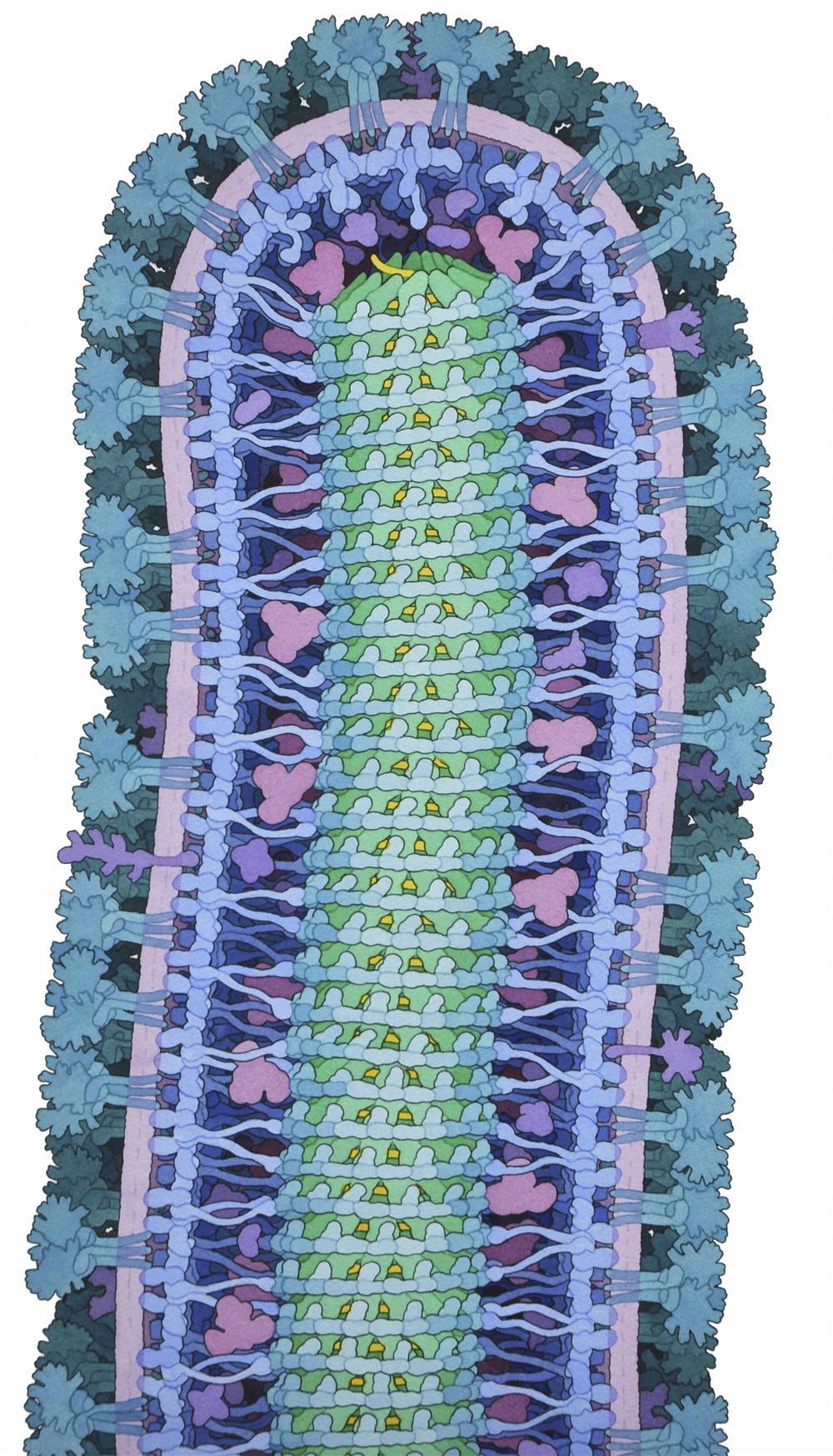
Ebola Virus
Photograph from 2014 of an ink and watercolor illustration depicting a cross section of an Ebola virus particle, appearing surrounded by a pink to purple membrane. The Ebola virus, which first appeared in Africa in the mid-1970s, spreads between humans through direct contact with infected blood, secretions and organs, and contaminated surfaces or bedding impregnated with these fluids. The image has been the winner of this edition.
Photo: David S. Goodsell, Protein Data Bank RCSB / Wellcome Images
Heart of Adult Cow
Photograph from 2013 showing the heart of an adult cow. The heart, 27 centimeters high, has four chambers through which blood flows. First it enters through the two upper cavities and then it exits through the two lower cavities.
Photo: Michael Frank, Royal Veterinary College / Wellcome Images
Corn leaves
Cross section of a set of corn leaves, one of the most cultivated cereals in the world, used as a staple food, livestock feed or to make high fructose corn syrup, popular in the United States as a sweetener for processed foods and beverages. The photograph, from 2013, was created in collaboration with Jim Haseloff.
Photo: Fernán Federici / Wellcome Images
Stem Cell
At the center of this 2015 image is a human stem cell embedded in a 3D porous hydrogel matrix, designed to mimic the real-world environment of the cell in the bone marrow.
Photo: Sílvia A Ferreira, Cristina Lopo and Eileen Gentleman, KCL / Wellcome Images
Parasites

Image from 2015 showing three tachyzoites, a developmental stage of Toxoplasma gondii, which is a species of parasitic protozoan that causes toxoplasmosis, a common infection in birds and mammals.
Photo: Leandro Lemgruber, University of Glasgow / Wellcome Images
Zebrafish Embryo
Photograph from 2015 showing asymmetric cell division in a living zebrafish brain embryo.
Photo: Paula Alexandre, UCL / Wellcome Images
Clatrin cage
Molecular model from X-ray diffraction data of a clathrin cage, a type of protein found in the liver and in other peripheral tissues. The photograph is from 2014.
Photo: RCSB Protein Data Bank / Wellcome Images
Bone Development
Image from 2013 showing bone development in the vertebrae of a baby's spine.
Photo: Frank Acquaah / Wellcome Images
Healthy Brain
2015 image of the healthy brain of a young adult. The brain is seen from behind.
Photo: Alfred Anwander, MPI-CBS/Wellcome Images
Nanographene oxide
Nanographene oxide (the purple background) interacts with two rod-shaped bacteria, in an image from 2015.
Photo: Izzat Suffian, Kuo-Ching Mei, Houmam Kafa & Khuloud T. Al-Jamal/ Wellcome Images
Swallowtail Butterfly
2014 photo of the head of a swallowtail butterfly. The two compound eyes, the two antennae and a long, curly trunk that it uses to extract nectar from flowers are clearly distinguishable.
Photo: Macroscopic Solutions/ Wellcome Images
Madagascar Moth
2015 image showing the scales of a Chrysiridia rhipheus moth during a sunset in Madagascar. Often mistaken for a butterfly, this moth flies during the day, while most moths fly at night.
Photo: Macroscopic Solutions/ Wellcome Images
Human liver tissue
2015 image of human liver tissue implanted into a mouse model of liver injury.
Photo: Chelsea Fortin, Kelly Stevens and Sangeeta Bhatia, Koch Institute, copyright MIT / Wellcome Images
Infectious Disease Containment Unit
One of the infectious disease containment units at the Royal Free Hospital in London. The unit was used in 2014 to contain a patient who was infected with the Ebola virus, which she contracted while working as a nurse in Sierra Leone.
Photo: Wellcome Images
Raynaud's disease
Image created in 2015 using thermal infrared, which allows you to see thermal energy or radiation, commonly known as heat. The image shows the hand of a person experiencing symptoms of Raynaud's disease (on the right) versus one who is not affected (on the left). The unaffected hand is seen to heat up much faster and begin to emit higher levels of radiation soon after exposure, while the affected hand continues to emit lower levels.
Photo: Thermal Vision Research / Wellcome Images
Retina of an eye
Three-dimensional image of the center of the retina of the eye of a healthy human.
Photo: Peter Maloca/ Wellcome Images
Allergic Reaction
Photograph from 2014 showing an allergic reaction to the agent paraphenylenediamine in a henna tattoo, causing skin blistering.
Photo: Nicola Kelley, Cardiff and Vale University Hospital NHS Trust / Wellcome Images
Occlusion of a vein
Central retinal vein occlusion in a photograph taken in 2015.
Photo: Kim Baxter, Cambridge University Hospitals NHS Foundation Trust / Wellcome Images
Premature Baby
Premature baby receiving UV phototherapy, a treatment that uses ultraviolet light. Some premature babies may have abnormal levels of bilirubin in their blood, which prevents their liver from functioning normally and that is why this treatment is performed. The photograph is from 2015.
Photo: David Bishop, Royal Free Hospital London / Wellcome Images
Stroke
Photograph from 2015 of stroke-related disability in adults.
Photo: Nicholas Evans, University of Cambridge / Wellcome Images
Last year, the winner was a photograph showing the uterus of a mare, with the fetus attached to the mother by the umbilical cord. Among the photographs selected in this edition there is an adult cow's heart, the healthy brain of a young man, the head of a swallowtail butterfly or the center of the retina of a human eye. The images will be exhibited at the Science Museum in London and in countries including the United States, Russia and South Africa.


Related Articles